
 Replacing a missing tooth sounds straightforward on paper, but the reality is that implant placement demands a level of precision most patients never think about. The jaw contains nerves, blood vessels, varying bone density, and neighboring roots that all need to be accounted for before a single drill contacts the bone. When clinicians rely on traditional methods, they work largely from two-dimensional X-rays and experience alone. That gap between what they can see and what is actually happening beneath the surface is exactly where complications tend to originate.
At Spring St. Dental in Bastrop, we take a different approach to dental implants by using CT-guided implant surgery, a technology-driven process that maps your anatomy in three dimensions before treatment begins. Patients who have had implants placed years ago and then experienced our guided process often describe it as a completely different procedure, one that feels more controlled, more predictable, and quicker to heal.
What CT-Guided Implant Surgery Actually Involves
Guided implant placement starts long before the day of your procedure. The process is rooted in the detailed three-dimensional data we collect using our in-office cone beam CT scanner, not the flat X-ray images that most patients associate with dental care.
The Role of CBCT Scanning
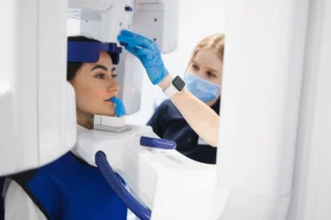
Our 3D cone beam CT produces a full volumetric image of your jaw, including the precise location of nerves, the depth and density of available bone, and the architecture of surrounding structures. Traditional two-dimensional X-rays compress all of that information into a flat image. When only a flat image is available, the clinician must estimate distances and angles that can only be fully understood in three dimensions.
With CBCT data, we can see exactly how much bone is available at each proposed implant site, where the inferior alveolar nerve runs, and whether bone grafting may be needed before placement. That information changes not just how the procedure goes but whether it should proceed the same way at all.
Building the Surgical Guide
Once we complete the 3D scan, we use that data to digitally plan the exact trajectory, depth, and angulation of each implant. That plan is then converted into a custom surgical guide, manufactured in-office using our 3D printer. The guide fits over your teeth or gums during the procedure and physically constrains each drill to follow the pre-planned path. Research published in PubMed Central examining computer-guided implant placement using CBCT found that guided techniques produced highly predictable positional accuracy between planned and placed implants, reinforcing why this workflow has become the standard of care for precision-focused practices.
How the Process Differs from Freehand Placement
In freehand implant placement, the clinician directs each drill based on visual landmarks, tactile feedback, and clinical judgment. Those are meaningful inputs, but they do not account for the three-dimensional variation that exists in every patient’s jaw. The guide removes that variability.
When the surgical guide is in place, the drill moves through a precisely sized sleeve that limits lateral movement. Depth stops prevent the drill from advancing beyond the planned endpoint. The result is that the implant is placed where the virtual plan says it should be, not where the clinician’s hand approximates it to be. For patients with limited bone volume, close proximity to a nerve, or complex restorative plans like a full arch restoration, that level of control is what separates a predictable outcome from a risky one.
There are several reasons why we perform all implants using CT-guided surgery rather than reserving it only for complex cases. These include:
- Consistent placement accuracy regardless of case difficulty
- Reduced surgical time because the path is predetermined
- Quicker recovery for most patients due to more controlled tissue management
- Greater confidence in final crown positioning, including fit with crowns and bridges that rely on precise implant angulation
Each of these factors contributes to outcomes that are more predictable from both a surgical and a restorative standpoint.
Why We Use This Approach for Every Implant Case
Most dental offices reserve guided implant surgery for cases they categorize as high-risk. We use it for every implant case because the information CBCT provides is valuable across the full spectrum of patient anatomy, not just the outliers. The diagnostic advantages of digital imaging extend well beyond the placement day: they inform bone quality assessments, identify issues that two-dimensional films would miss, and give patients a clearer picture of what the procedure involves before they commit to it.
There is also a practical benefit for recovery. Because the surgical guide directs the procedure, we can often complete placement with less disruption to surrounding tissue than a freehand approach would require. For many patients, that translates to a quicker and more comfortable post-operative experience.
Begin Your Implant Consultation at Spring St. Dental
Spring St. Dental brings together Dr. Brian Romney, DMD, Dr. Matthew Lassen, DMD, and Dr. Meghan Bhakta, DDS, each with distinct clinical backgrounds and a shared commitment to technology-driven, patient-centered care. We perform all implant procedures in-office using our CBCT scanner and in-house 3D printing capability. That combination allows us to plan, fabricate, and place each implant within a controlled, fully digital workflow.
If you are considering an implant or have questions about whether guided placement is right for your situation, we are here to walk you through it. Contact us to schedule a consultation with our team.