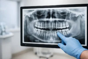
 A tooth in distress doesn’t announce itself with a clear roadmap. The canals hidden inside each root are narrow, curved, and sometimes branching in ways that a flat X-ray simply cannot capture, and what a dentist can’t see can absolutely impact the outcome of treatment. Three-dimensional imaging has changed what’s possible in root canal therapy, giving clinicians a level of anatomical insight that two-dimensional radiographs were never designed to provide.
At Spring St. Dental in Bastrop, Texas, the team embraces technology as a core part of delivering thorough, precise care. Root canal therapy performed with advanced imaging support enables the team to assess the root structure, identify potential complications, and plan treatment with confidence, benefiting every patient. Whether you’re coming in with persistent tooth pain or a referral for endodontic treatment, understanding what 3D imaging brings to the process helps set accurate expectations.
What Traditional X-Rays Miss
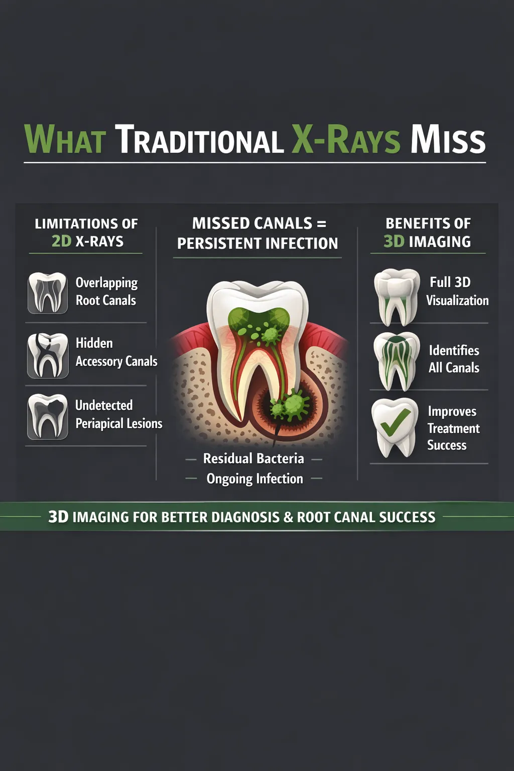
Conventional two-dimensional radiographs have long been the standard in dental diagnosis, and they remain useful in many clinical settings. However, they compress a three-dimensional structure into a flat image, which creates inherent blind spots. Root canals that overlap one another on a 2D image, accessory canals that branch off unexpectedly, and the true extent of periapical lesions can all be obscured or misrepresented in standard imaging.
This matters during a root canal because incomplete treatment, often resulting from canals missed or anatomy misread, is one of the primary reasons procedures fail. When a canal goes undetected and untreated, bacteria remain, and infection can persist or return. This is not a failure of the procedure itself but often a diagnostic limitation that 3D imaging directly addresses.
How CBCT Scanning Supports Endodontic Diagnosis
Cone beam computed tomography, commonly called CBCT, generates a three-dimensional reconstruction of the tooth and surrounding bone from a single scan. Unlike traditional CT used in medical settings, dental CBCT is calibrated for the oral and maxillofacial region, delivering detailed imagery at a radiation dose suited to the diagnostic task at hand.
According to a 2025 systematic review published in the National Library of Medicine, research on 3D imaging in endodontics has consistently shown that CBCT provides more comprehensive visualization of root canal anatomy than two-dimensional imaging, including clearer views of canal curvature, number, and form. This level of detail is especially valuable for teeth with complex anatomy, such as molars with multiple roots or canals following irregular paths.
What CBCT Reveals Before Treatment Begins
Before a root canal begins, a CBCT scan can show the treating dentist the exact number and orientation of canals, the degree of root curvature, any signs of resorption or fracture, and the proximity of the tooth to adjacent anatomical structures, such as nerve pathways or the sinus cavity. This pre-treatment clarity allows for more accurate planning and reduces the likelihood of surprises during the procedure. Understanding what is involved in endodontics before treatment begins helps patients engage meaningfully in the process and make informed decisions about their care.
Recognizing When 3D Imaging Is Indicated
Not every root canal requires a CBCT scan, and clinical judgment guides when 3D imaging provides meaningful additional value. Cases where 3D imaging is most beneficial include those involving symptoms suggestive of complex or advanced infection, retreatment scenarios in which previous procedures complicate the anatomy, suspected root fractures, unusual pain presentations, or teeth with atypical canal configurations on standard imaging. When the 2D picture raises more questions than it answers, a 3D scan is the logical next step.
Improved Accuracy During Treatment
The value of 3D imaging doesn’t end at diagnosis. For clinicians, having a pre-treatment CBCT available during the procedure means working from a more complete anatomical reference. This supports more precise access cavity preparation, reduces unnecessary removal of healthy tooth structure, and helps ensure each identified canal is properly cleaned and shaped.
It also supports better verification after treatment. Follow-up imaging can confirm that the root filling material is properly placed throughout the canal system and that the periapical area is healing appropriately. For patients who have had previous endodontic work and are presenting with lingering discomfort, reviewing what diagnostic imaging can uncover provides important context. The result is a more informed clinician and a more confident patient.
How This Fits Into Preventive and Restorative Care
Root canal therapy doesn’t exist in isolation. It is often the step that preserves a tooth that would otherwise require extraction, allowing patients to maintain natural dentition and avoid the downstream need for implants or prostheses. When a tooth can be saved with precision endodontic treatment, it supports long-term oral health in a meaningful way. The commitment to thorough preventive dentistry and restorative care at Spring St. Dental reflects the understanding that keeping natural teeth healthy is always the preferred outcome.
The integration of 3D imaging into root canal therapy represents a thoughtful, evidence-based approach producing better outcomes, fewer missed diagnoses, and a smoother experience for patients already dealing with significant dental discomfort. It is a tool supporting clinical decision-making at every stage of the process.
Schedule Your Visit With Spring St. Dental
Spring St. Dental is committed to delivering thorough, precise dental care in a calm and relaxed environment. The team works to ensure every patient receives individualized attention, accurate diagnosis, and a clear understanding of treatment options before any procedure begins. With a philosophy rooted in advanced, conservative techniques and a dedication to preserving natural teeth wherever possible, patients across Bastrop and the surrounding communities receive care meeting a high standard.
If you’re experiencing tooth pain, have been told you may need a root canal, or want to discuss your dental health with a team taking the time to listen, reaching out is the first step. Contact us to request an appointment and connect with the Spring St. Dental team today.